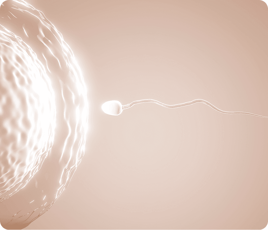
In vitro
Skuteczny zabieg zapłodnienia metodą in vitro po raz pierwszy przeprowadzono w 1977 r. u Pacjentki leczącej się z powodu niedrożnych jajowodów. Z biegiem lat i udoskonalaniem metody rozszerzyła się lista wskazań kwalifikujących Pacjentów do zabiegu, jednocześnie zminimalizowano częstotliwość występowania powikłań i działań niepożądanych.
Współcześnie zapłodnienie pozaustrojowe jest coraz częściej rekomendowaną metodą leczenia o najwyższej skuteczności ze wszystkich dostępnych technik wspomaganego rozrodu.
Co to jest in vitro?
Zapłodnienie pozaustrojowe (łac. in vitro, dosłownie: „w szkle”) to procedura polegająca na połączeniu w warunkach laboratoryjnych, uprzednio pobranych i odpowiednio wypreparowanych, żeńskich i męskich komórek rozrodczych oraz na przeniesieniu uzyskanych zarodków do macicy Pacjentki.
-
Zapłodnienie pozaustrojowe uznawane jest za metodę leczenia niepłodności i klasyfikowane jako jedna z technik wspomaganego rozrodu (ART, ang. assisted reproductive technology). Techniki te to metody pozwalające na uzyskanie ciąży z pomięciem co najmniej jednego etapu poczęcia przebiegającego w warunkach fizjologicznych.
-
Moment połączenia w laboratorium przez embriologa plemnika z komórką jajową poprzedza stymulacja hormonalna Pacjentki (wyjątek: Pacjentki podchodzące do procedury na tzw. „własnym cyklu”), punkcja jajników (pobranie płynu pęcherzykowego, w którym zawieszone są komórki jajowe) oraz preparatyka nasienia. Uzyskane w efekcie zapłodnienia metodą in vitro zarodki są następnie hodowane w laboratorium, (optymalnie) jeden transferowany jest do macicy Pacjentki, a pozostałe – kriokonserwowane do późniejszego wykorzystania podczas kriotransferów.
-
Skuteczny zabieg zapłodnienia metodą in vitro po raz pierwszy przeprowadzono w 1977 r. u Pacjentki leczącej się z powodu niedrożnych jajowodów. Z biegiem lat i udoskonalaniem metody rozszerzyła się lista wskazań kwalifikujących Pacjentów do zabiegu, jednocześnie zminimalizowano częstotliwość występowania powikłań i działań niepożądanych. Współcześnie zapłodnienie pozaustrojowe jest coraz częściej rekomendowaną metodą leczenia o najwyższej skuteczności ze wszystkich dostępnych technik wspomaganego rozrodu.

Przygotowanie do in vitro – kobieta
Zachęcany do obejrzenia filmu, w którym znajdują się informacje na temat przygotowania kobiety do in vitro oraz przebiegu tej procedury.

Wskazania do in vitro – mężczyzna i kobieta
Zachęcany do obejrzenia filmu, w którym znajdują się informacje na temat wskazań dla mężczyzn przygotowujących się do in vitro.
Szczegółowy opis
Poniżej przedstawiamy listę najczęstszych czynników wpływających na płodność, które diagnozujemy i leczymy.
brak lub nieoperacyjna niedrożność jajowodów. Zmiany w jajowodach i okolicach przydatków, które uniemożliwiają lub utrudniają migrację plemników oraz przemieszczanie się zapłodnionej komórki i zarodka przez jajowody:
- u Pacjentek z trwałym uszkodzeniem jajowodów,
- u Pacjentek wykluczonych z leczenia operacyjnego mającego na celu odbudowanie prawidłowej funkcji jajowodów,
- u Pacjentek z upośledzoną funkcją jajowodów przy zachowanej drożności lub po operacji mikrochirurgicznej i upływie 2. lat bez ciąży.
kiedy żadne z przeprowadzonych badań diagnostycznych nie wskazuje przyczyny niezamierzonej bezdzietności:
- jeżeli niepłodność trwa dłużej niż 3 lata (2 lata wg NICE),
- jeżeli Pacjentka ma ukończone 35 lat, przyjmuje się krótszy okres trwania niepłodności
Endometrioza to przewlekła choroba ginekologiczna dotycząca 5-10% kobiet w okresie reprodukcyjnym, prowadząca do długotrwałego stanu zapalnego. Charakteryzuje się występowaniem ognisk histopatologicznie podobnych do błony śluzowej macicy, ale poza macicą (zwłaszcza na otrzewnej miednicy małej) powodujących zrosty i niedrożność jajowodów, na jajnikach – powodujących torbiele i gorszą jakość oocytów, na przegrodzie odbytniczo-pochwowej – bóle podczas współżycia.
Występowanie tkanek zbliżonych budową do endometrium poza jamą macicy, skutkujące zmianami mechanicznymi, zapalnymi, wysiękami krwi i następowymi zrostami w obrębie układu rozrodczego lub poza nim:
- I, II stopień, po okresie naturalnych starań, takim jak w przypadku niepłodności niewyjaśnionego pochodzenia,
- III, IV stopień.
12 cykli stymulowanych bez uzyskania ciąży.
Co dotyczy kobiet:
- z usuniętymi jajnikami (np. z przyczyn nowotworowych lub zapalnych),
- z przedwczesną menopauzą,
- po wielu nieudanych zabiegach z własnymi komórkami jajowymi,
- wyniki badań wskazują na wyczerpującą się liczbę komórek jajowych.
(badania hormonalne wskazują, że zachowany został proces spermatogenezy)
w zależności od potencjału rozrodczego kobiety wskazaniem jest inseminacja lub zabieg zapłodnienia in vitro z użyciem nasienia dawcy.
Stymulacja hormonalna ma na celu kontrolowane pobudzenie jajników do produkcji pęcherzyków jajnikowych zawierających komórki jajowe. Podczas jednego cyklu powstaje od kilku do kilkunastu takich pęcherzyków.
W większości przypadków w cyklu poprzedzającym rozpoczęcie procesu stymulacji jajników Pacjentka przyjmuje tabletki antykoncepcyjne. Ma to na celu wyciszenie pracy jajników i przeciwdziałanie tworzeniu się ich torbieli. Pod koniec tego cyklu, na wizycie lekarskiej połączonej z badaniem USG, zapada ostateczna decyzja co do rozpoczęcia i rodzaju stymulacji hormonalnej. W zależności od wieku Pacjentki, poziomu hormonów w surowicy krwi, przebiegu dotychczasowego leczenia oraz obrazu jajników w USG lekarz zaleca jeden z trzech schematów: protokół długi, krótki lub krótki z antagonistą.
Para przyjeżdża na zabieg ok. 15. minut przed wyznaczoną godziną punkcji. Zabieg poprzedza rozmowa z anestezjologiem, w trakcie której lekarz zapoznaje się z najbardziej aktualnym stanem zdrowia Pacjentki oraz zakłada do żyły plastikową igłę (venflon), służącą następnie do podaży leków i przetaczania płynów. Venflon zostanie usunięty przez pielęgniarkę w gabinecie zabiegowym tuż przed opuszczeniem kliniki przez Pacjentkę, po kontroli ciśnienia krwi, czyli około 3 – 6. godzin po punkcji.
Bezpośrednio po pobraniu płyn pęcherzykowy pobrany trafia do laboratorium IVF, gdzie pod mikroskopem wyszukiwane są komórki jajowe. Komórka jajowa ma ok. 0,2 mm średnicy. Wraz z otaczającym ją wzgórkiem jajonośnym, czyli warstwą komórek ziarnistych odżywiających komórką jajową, jest dobrze widoczna gołym okiem. Znalezione komórki przenosi się do specjalnych odżywek i umieszcza w inkubatorze.
W tym czasie przygotowuje się oddane do specjalnego pojemnika i dostarczone do laboratorium nasienie Partnera. Przygotowanie polega na wyselekcjonowaniu w odpowiedniej pożywce i przy zastosowaniu technik wirowniczych najlepiej zbudowanych i najżywotniejszych i plemników.
Transfer zarodków następuje rutynowo w drugiej lub trzeciej dobie po punkcji. Zarodek na tym etapie ma cztery do ośmiu komórek. W cienkim plastikowym cewniku umieszcza się maksymalnie dwa zarodki i wraz z małą ilością (0,3 ml) płynu hodowlanego podaje do macicy. Zabieg odbywa się na fotelu ginekologicznym, w gabinecie transferowym, który sąsiaduje z laboratorium IVF. Jest bezbolesny i nie wymaga znieczulenia. Podanie zarodka przeprowadza się przy wypełnionym pęcherzu moczowym.
Jeśli test potwierdza ciążę, Pacjentka kontynuuje leczenie progesteronem aż do zakończenia I trymestru ciąży (10 – 12 tygodni od przeprowadzenia zabiegu IVF) – w tym czasie dawka leku zostanie skorygowana w zależności od aktualnych wyników badań. Badanie USG potwierdzające ciążę wykonuje się dwukrotnie: w 25. dniu po zabiegu w celu ustalenia czy pęcherzyk płodowy znajduje się macicy, a kolejne, w 35. dniu po transferze, w celu zarejestrowania czynności serca płodu. Jeśli badanie wykonywane jest w nOvum, jest to ostatnia wizyta u lekarza prowadzącego dotychczasowe leczenie.
Wszystkie rokujące zarodki, które powstają w laboratorium in vitro i nie zostaną podane do macicy, zostają zamrożone do późniejszego wykorzystania. Możliwość wykorzystania zamrożonych zarodków pozwala uniknąć stymulacji hormonalnej i punkcji jajników u pacjentki ponownie starającej się o ciążę po urodzeniu dziecka lub po nieudanym pierwszym transferze – oszczędza więc zdrowie kobiety oraz obniża koszty leczenia, dlatego jest bardzo ważnym etapem procedury zapłodnienia in vitro.
Zarodki przeznaczone do późniejszego wykorzystania, podobnie zresztą jak plemniki i komórki jajowe, poddawane są procesowi kriokonserwacji w ciekłym azocie. Jest to obecnie jedyna znana metoda umożliwiająca ich przeżycie.
Po porodzie lub kiedy próba uzyskania ciąży po transferze tzw. „świeżych” zarodków nie powiedzie się, w kolejnych cyklach Pacjentka może przystąpić do transferu zarodków poddanych wcześniej kriokonserwacji (kriotransfer). Nie ma wówczas potrzeby powtórnego przechodzenia przez wszystkie etapy stymulacji i pełnego przygotowania do zabiegu zapłodnienia pozaustrojowego.
Aby przystąpić do zabiegu kriotransferu, Pacjentka powinna umówić się na wizytę do lekarza prowadzącego bezpośrednio przed miesiączką, po której ma nastąpić kriotransfer lub najpóźniej do 5. dnia właściwego cyklu. Jeśli USG narządu rodnego wykonane w czasie wizyty nie wykaże żadnych nieprawidłowości oraz nie ma innych przeciwwskazań zdrowotnych, rozpoczyna się przygotowanie do kriotransferu.
Kriotransfer może być przeprowadzony w naturalnym cyklu owulacyjnym (jeżeli Pacjentka ma owulacje) lub w tzw. cyklu. bezowulacyjnym, w którym odpowiednią grubość śluzówki macicy osiąga się podając leki zawierające estrogeny.
Bezpieczne in vitro w nOvum
Jednym z najważniejszych oczekiwań Par leczących się z powodu niepłodności jest pewność jednoznacznej identyfikacji komórek i zarodków, które powierzają Laboratorium embriologicznemu oraz BKRiZ. Od momentu pobrania komórek jajowych i plemników kontrolę i opiekę nad nimi sprawują embriolodzy. Jest to ogromna odpowiedzialność, wymagająca najsprawniej działających systemów kontroli jakości oraz identyfikacji materiału na każdym etapie prac laboratoryjnych. Dotyczy to zarówno pobranych komórek przed zapłodnieniem, jak i identyfikacji prowadzonych hodowli zarodkowych, identyfikacji podczas transferu, przygotowania do kriokonserwacji, przechowywania i kriotransferów.
W nOvum od lat pracujemy zgodnie z autorskim systemem wielokrotnej kontroli, lecz jednocześnie śledzimy rozwój światowych technologii w tym zakresie. W 2015 roku podjęliśmy strategiczną decyzję o wprowadzeniu elektronicznego i niezależnego systemu kontroli pracy na każdym etapie działań Laboratorium embriologicznego nOvum – system RI Witness, działający w najlepszych światowych laboratoriach embriologicznych. Jest to znaczące przedsięwzięcie, które podjęliśmy mając na uwadze bezpieczeństwo prowadzonych w nOvum procedur, komfort Pacjentów powierzających nam swój materiał biologiczny oraz komfort pracy embriologów, którzy w większym stopniu mogą koncentrować się na pracy medycznej. Stały monitoring przebiegu procesów embriologicznych zapewniony przez system RI Witness pozwala zminimalizować ryzyko błędu ludzkiego związanego z realizacją powtarzalnych zadań i błędnym postrzeganiem.

System RI Witness
W OMWP i BKRiZ nOvum wprowadzony został RI Witness – unikalny system znakowania komórek rozrodczych i zarodków, gwarantujący bezbłędną identyfikację na etapie ich pobierania, każdym z etapów działań laboratoryjnych oraz etapie przechowywania.
RI Witness to elektroniczny i niezależny system kontroli pracy na każdym etapie działań Laboratorium seminologicznego, embriologicznego i BKRiZ oraz sali zabiegowej.
- Każda Para rozpoczynająca procedurę otrzymuje kartę ze swoim osobistym i unikalnym kodem – oznakowane dla każdego Pacjenta i Pacjentki indywidualne karty systemu RI Witness stanowią źródło do dalszego znakowania materiału biologicznego pobranego od Pacjentów na dalszych etapach leczenia.
- System RI Witness wykorzystywany jest na wszystkich etapach procedur przeprowadzanych w Laboratoriach seminologicznym, embriologicznym oraz BKRiZ nOvum i sali zabiegowej (pobieranie gamet, zapłodnienie pozaustrojowe, hodowla zarodkowa, transfer zarodka, mrożenie i rozmrażanie gamet oraz zarodków).
- Wszystkie naczynia wykorzystywane do pobierania i przenoszenia materiału opatrzone są tym samym kodem – znakowanie naczyń (pojemniki, probówki, szalki itp.) odbywa się łańcuchowo zaczynając od kart zakodowanych na konkretnego Pacjenta/Pacjentkę.
- Na każdym etapie procesu zapłodnienia pozaustrojowego, znaczniki ID oraz identyfikacja kodu przez system elektroniczny (technologia RFID) pozwalają na identyfikowanie, śledzenie i prowadzenie rejestru materiału biologicznego pacjentów.
- Stanowiska pracy w Laboratoriach seminologicznym i embriologicznym oraz BKRiZ i sali zabiegowej odbiera bezprzewodowy sygnał wysyłany przez znaczniki ID, zapewniając bezpieczny rejestr całego cyklu. Oznacza to, że każdy etap jest monitorowany i rejestrowany przez system niezależny od wewnętrznych procedur naszych laboratoriów.
- RI Witness wielokrotnie potwierdza tożsamość materiału biologicznego i dopiero po potwierdzeniu dopuszcza do dalszego etapu pracy i działań laboratoryjnych.
- W (mało prawdopodobnym) przypadku, gdyby system zgłosił wątpliwości co do tożsamości komórek lub zarodków, system automatycznie zablokuje proces laboratoryjny, zapobiegając potencjalnej pomyłce. W takiej sytuacji wdrażana jest procedura wyjaśniająca.
Przez wprowadzenie przez nas w OMWP i BKRiZ systemu RI Witness otrzymujecie Państwo gwarancję, że:
- każdy z etapów składających się na proces leczenia technikami wspomaganego rozrodu w OMWP i BKRiZ nOvum jest bezpiecznie monitorowany;
- na etapie pobrania gamet i każdym etapie procedur laboratoryjnych materiał biologiczny jest jednoznacznie i bez żadnych wątpliwości identyfikowany i zarazem identyfikowalny;
- leczycie się Państwo w Klinice, gdzie z sukcesem przeprowadza się znaczną liczbę procedur zapłodnienia pozaustrojowego, a Państwa bezpieczeństwo i efekt leczenia są dla nas priorytetem. Filozofia nOvum opiera się na zachowaniu najwyższych standardów, kontroli jakości, a każdy etap leczenia jest w pełni nadzorowany;
- Państwa komórkami rozrodczymi i zarodkami opiekują się embriolodzy pracujący w komfortowych warunkach, pozwalających na wykonywanie kolejnych etapów procedur z pełną koncentracją i uwagą;
- Państwa komórki rozrodcze i zarodki znajdują się w bezpiecznym miejscu pod ścisłym nadzorem embriologów, kriobiologów i sprawnie funkcjonujących systemów elektronicznych;
- przez lata przechowywania zarodki i komórki rozrodcze są identyfikowalne według unikalnego systemu – są bezpieczne!
Zabiegi wspomagające skuteczność in-vitro
Docytoplazmatyczna iniekcja plemnika do wnętrza komórki jajowej (ICSI; ang. intracytoplasmic sperm injection) to wysokospecjalistyczna technika wspomaganego rozrodu – metoda zapłodnienia pozaustrojowego, w której pojedynczy plemnik wstrzykuje się bezpośrednio do wnętrza komórki jajowej za pomocą szklanej igły, tzw. mikropipety.
Wskazania do zastosowania ICSI obejmują sytuacje, gdy:
- w nasieniu mężczyzny stwierdza się zbyt małą liczbę plemników lub też ich ruchliwość jest nieprawidłowa;
- wykonane w przeszłości próby zapłodnienia pozaustrojowego kończyły się niepowodzeniem (nie dochodziło do zapłodnienia);
- pobrano mniej niż 5 komórek jajowych w czasie punkcji;
- Pacjentka ma endometriozę lub niepłodność o niewyjaśnionej przyczynie (idiopatyczną).
Jedną z odmian technik zapłodnienia metodą mikromanipulacji jest tzw. fizjologiczne ICSI – pICSI (ang. phisiological ICSI). Na główkach plemników, które prawidłowo przeszyły proces spermatogenezy obecne są specjalne receptory, dzięki którym męskie gamety wchodzą w reakcje z hialuronianem macierzy wzgórka jajonośnego oocytu. Taki ejakulat określany jest jako biologicznie dojrzały, a plemniki gotowe do skutecznego zapłodnienia komórek jajowych.
W warunkach laboratoryjnych reakcja plemnika z kwasem hialuronowym pozwala embriologowi ocenić jego jakość – plemniki wykazujące zdolność wiązania z kwasem hialuronowym są w pełni dojrzałe i z powodzeniem mogą być użyte do zapłodnienia komórek jajowych metodą pICSI. Zastosowanie metody pICSI zwiększa szansę na uzyskanie większej liczby wysokiej jakości zarodków, a co za tym idzie – szans na pomyślną implantację i ciążę.
W przypadku całkowitego braku plemników w nasieniu, czyli azoospermii, istnieje możliwość wykonania biopsji najądrza lub jądra u mężczyzny i, jeśli znajdą się tam żywe plemniki, wykonania mikroiniekcji takich plemników do wnętrza komórek jajowych. Metody te, zwane w angielskich skrótach PESA i TESE, M-TESE, pozwalają na ojcostwo w sytuacjach, w których jeszcze niedawno nie było to możliwe.
PESA (ang. Percutaneous Epididymal Sperm Aspiration) to przezskórna aspiracja plemników z najądrza.
TESE (ang. Testicular Sperm Extraction) to ekstrakcja plemników z jądra.
M-TESE (ang. Micro – Testicular Sperm Extraction) to pobranie fragmentów tkanki w trakcie otwartej biopsji jąder, kontrolowanej pod mikroskopem operacyjnym, w celu uzyskania plemników do zapłodnienia in vitro.
Pobrana w czasie zabiegu próbka bioptatu/aspiratu z najądrzy jest oglądana pod mikroskopem w poszukiwaniu plemników, druga próbka jest wysyłana do badania histopatologicznego. Pozostały materiał – niezależnie od tego czy w próbce znalezione zostały plemniki, czy nie – jest zamrażany, aby mieć wiedzę czy jest szansa na znalezienie plemników i czy rozpoczęcie stymulacji jajników jest zasadne. Gdy wynik badania histopatologicznego potwierdza szansę na znalezienie plemników, Pacjentka jest kwalifikowana do stymulacji i zabiegu zapłodnienia in vitro.
W dniu punkcji jajników i po uzyskaniu komórek jajowych, rozmrażane są kolejno poszczególne próbki aspiratu/bioptatu w poszukiwaniu żywych plemników zdolnych do zapłodnienia.
Wykonując biopsję igłową przezskórną lub otwartą najądrzy (PESA, MESA) lub jąder (TESA, TESE), pobiera się materiał, który przekazywany jest do laboratorium IVF. Tam, pod mikroskopem, wyszukiwane są plemniki, które mogą być użyte w protokole IVF-ICSI. Jeśli w pobranej tkance plemniki się nie znajdą, biopsję można powtarzać – nawet wielokrotnie – jednak nie praktykuje się tego, ponieważ jest to istotne obciążenie dla pacjenta, a ponadto szansa na znalezienie plemników w kolejnych pobraniach (>= 3) spada praktycznie do zera.
W przypadku M-TESE tkankę z jąder pobiera się pod kontrolą mikroskopu w taki sposób, aby bardzo precyzyjnie określić miejsca, z których zostaną wycięte kanaliki nasienne rokujące obecność spermatogenezy. Dlatego też M-TESE wielokrotnie przewyższa precyzją zabiegi PESA, MESA, TESA czy TESE.
IMSI (ang. intracytoplasmic morphologically selected sperm) to jedna z odmian zapłodnienia pozaustrojowego metodą mikromianipulacji. W przypadku tradycyjnej metody zapłodnienia pozaustrojowego w procedurze ICSI, stosowane przy wyborze plemnika do zapłodnienia powiększenie obrazu spod mikroskopu jest na tyle małe (400 razy), że nie pozwala uwidocznić wyraźnie wszystkich szczegółów jego budowy.
W procedurze IMSI zastosowanie optycznego powiększenia rzędu 600 – 1000 razy przy jednoczesnym dodatkowym powiększeniu cyfrowym, pozwala finalnie uzyskać powiększenie obrazu rzędu 6000 – 10000 razy, co umożliwia dokładniejsze obejrzenie plemników i wybranie tych o najlepszej morfologii, co ponoć pozwala podnieść wskaźnik skuteczności zapłodnienia pozaustrojowego.
Metoda IMSI została wprowadzona przez B. Bartoove’a w 2001 roku. Według zaleceń Bartoove’a ocenie podlegać powinny: akrosom, jądro, region zaakrosomalny, wstawka, mitochondria, witka. Najważniejszą częścią plemnika, na której skupia się twórca metody we wszystkich doniesieniach naukowych, jest główka plemnika, a w szczególności jej część zawierająca jądro komórkowe. Prawidłowy plemnik powinien mieć następujące wymiary: długość główki – 4,75 (+/- 0,28) μm, szerokość główki – 3,28 (+/- 0,20) μm, brak innych nieprawidłowości dotyczących innych struktur plemnika. W okolicy jądrowej główki plemnika nie powinno być więcej niż jednej wakuoli o wielkości poniżej 0.78±0.18 μm, zajmującej poniżej 4% powierzchni jądra. Od obecności wakuoli w okolicy jądrowej główki plemnika, które według Bartoove’a mają świadczyć o fragmentacji DNA, zależeć ma prawidłowy rozwój zarodka i szansa na prawidłowy rozwój ciąży.
Nie ma ogólnie przyjętych wskazań do IMSI, ponieważ nie ma udowodnionej wyższej skuteczności IMSI nad ICSI. Za wskazania do IMSI w nOvum przyjmuje się:
- powtarzające się niepowodzenia w zagnieżdżeniu zarodka po ICSI;
- poronienia po ICSI po wykluczeniu innych możliwych przyczyn tego stanu rzeczy;
- brak blastocyst lub niski odsetek blastocyst w przedłużonej hodowli in vitro;
- niepłodność męską w połączeniu w niskim odsetkiem prawidłowych plemników.
Procedura IMSI jest procedurą wysokospecjalistyczną i bardzo czasochłonną, co nieraz może mieć niekorzystny wpływ na komórki jajowe oczekujące na szczegółową ocenę plemnika, zanim zostaną zapłodnione. Także obraz plemników pod powiększeniem 1000-krotnym jest nieraz trudny do interpretacji, ponieważ niektóre dotychczas uznane za nieprawidłowe zmiany w ich budowie przez innych są interpretowane jako zmiany fizjologiczne.
Dojrzewanie komórek in vitro (IVM, ang. In Vitro Maturation) w laboratorium stosuje się u kobiet, dla których ze względu na budowę jajników, stymulacja hormonalna mogłaby być niebezpieczna poprzez powodowanie powstania nadmiaru pęcherzyków i ryzyko przestymulowania.
U takich Pacjentek wcześniej niż zwykle pobiera się komórki z niestymulowanych jajników lub stymulowanych minimalnymi dawkami leków. Pobrane komórki są niedojrzałe, niezdolne do zapłodnienia. Umieszcza się je w odżywkach zawierających hormony, które w klasycznym zapłodnieniu in vitro są podawane kobiecie do stymulacji. W wyniku tego, następnego dnia po punkcji (od 28. do 32. godzin dojrzewania w inkubatorze) ok. 40 – 70% komórek jajowych staje się zdolnych do zapłodnienia metodą ICSI. Po zapłodnieniu dalsze etapy leczenia przebiegają podobnie jak w klasycznym zapłodnieniu in vitro. Szanse na ciążę są o ok. 10 – 20% mniejsze, ale mniejsze jest też ryzyko powikłań w przypadku predysponowanych do niego Pacjentek.
Nie wszystkie cykle są odpowiednie, aby przeprowadzić punkcję, pobranie i dojrzewanie komórek jajowych. Ostateczna kwalifikacja następuje w 3. dniu cyklu na podstawie poziomu estradiolu w surowicy i badania USG.
Przygotowanie do zabiegu IVM polega na monitorowaniu cyklu i obserwacji wzrastających pęcherzyków Graafa, a także badaniu poziomu estradiolu w surowicy. Punkcja jest przeprowadzana między 7. a 10. dniem cyklu, kiedy pęcherzyki, endometrium i poziom estradiolu osiągną odpowiednią wielkość. W dniu punkcji konieczne jest pobranie 10. ml krwi w celu wypreparowania surowicy potrzebnej do dodania do medium hodowlanego.
Genetyczne badanie pierwszych ciałek kierunkowych w komórkach jajowych (PBB, ang. Polar Body Biopsy) zwiększa szansę na prawidłowo rozwijającą się ciążę i urodzenie zdrowego dziecka Parom decydującym się na zabieg zapłodnienia in vitro.
Ciałko kierunkowe jest to niewielki, zewnętrzny fragment komórki jajowej, powstający w wyniku jej dojrzewania, za pomocą którego komórka pozbywa się nadmiaru chromosomów. Na podstawie badania liczby chromosomów znajdujących się w ciałku kierunkowym można, z dużym prawdopodobieństwem, ocenić liczbę chromosomów w komórce jajowej.
Każda komórka organizmu człowieka (z wyjątkiem dojrzałych komórek rozrodczych) posiada w warunkach prawidłowych 46 chromosomów (struktur zbudowanych z DNA, w których znajdują się geny). Dojrzałe komórki rozrodcze: komórki jajowe i plemniki, posiadają po 23 chromosomy tak, aby po ich połączeniu nowy organizm posiadał ich 46. Pierwotnie 46-chromosomowa komórka jajowa dojrzewając, podczas tzw. podziału mejotycznego, tworzy ciałka kierunkowe (pierwsze i drugie) i za ich pomocą pozbywa się 23. chromosomów, robiąc miejsce na przyjęcie materiału genetycznego plemnika. W naturze, po spełnieniu tej ważnej funkcji, ciałka kierunkowe ulegają fragmentacji i zanikają. W warunkach laboratoryjnych można bez szkody dla komórki jajowej zbadać zawartość ciałka kierunkowego i na tej podstawie pośrednio ocenić materiał genetyczny matczynej komórki, ponieważ liczba chromosomów w pierwszym ciałku kierunkowym powinna odpowiadać liczbie chromosomów w komórce jajowej. Oczywiście, co należy podkreślić, przeprowadzone testy nie wykluczają wszystkich możliwych chorób genetycznych przenoszonych z organizmu matczynego, a jedynie z dużym prawdopodobieństwem wykluczają te najczęściej się pojawiające, związane z liczbowymi aberracjami chromosomowymi (nieprawidłową liczbą chromosomów).
Różnica między badaniem genetycznym ciałka kierunkowego niezapłodnionej komórki jajowej (PCGD), a diagnostyką przedimplantacyjną zarodka (PGD)
Diagnostyka przedimplantacyjna (PGD, ang. Preimplantation Genetic Diagnosis) to badanie istniejącego już zarodka pod kątem genetycznym. Jeśli zarodek okaże się nieprawidłowy, nie jest transferowany do macicy. Pozwala to uniknąć ciąży obciążonej genetycznie, nie przeciwdziała jednak powstawaniu wadliwych zarodków.
Dla części Pacjentów, którzy z powodu problemów medycznych mogą spodziewać się obciążeń genetycznych u potomstwa, technika diagnostyki przedimplantacyjnej PGD z etycznego punktu widzenia jest trudna do zaakceptowania. Selekcja istniejących już zarodków pod kątem wad genetycznych jest metodą stosowaną na świecie od lat, lecz nadal budzi kontrowersje, zarówno etyczne, jak i medyczne.
Szacunek dla takiego punktu widzenia naszych pacjentów skłonił nOvum do zastosowania diagnostyki chorób genetycznych na wcześniejszym etapie – badania niezapłodnionych komórek jajowych (PCGD, ang. Preconception Genetic Diagnosis) i zapładniania tylko tych, które okażą się prawidłowe pod względem liczby badanych chromosomów.
Wskazania do badania ciałka kierunkowego
Wskazaniem do wykonania badania jest uzasadnione podejrzenie wystąpienia u potomstwa trisomii chromosomów (obecność trzech zamiast dwóch chromosomów, np. zespół Downa, Edwardsa, Patau) lub monosomii (obecność jednego zamiast dwóch chromosomów), a także innych chorób genetycznych, których źródło leży w materiale genetycznym komórki jajowej. Wcześniej urodzone chore dzieci danej matki, wielokrotne niepowodzenia zabiegów zapłodnienia in vitro, poronienia, wiek kobiety powyżej 35. roku życia to powody do rozważenia zastosowania diagnostyki ciałka kierunkowego. Decyzja o opóźnianiu posiadania potomstwa nie zmienia twardych reguł biologii – u kobiet po 40. roku życia ok. 70% komórek jajowych jest nieprawidłowych pod względem liczby chromosomów.
Przebieg diagnostyki komórek jajowych
Komórki jajowe, po pobraniu z jajników, przenoszone są do laboratorium IVF. Z dojrzałych komórek jajowych pobiera się następnie ciałko kierunkowe, które jest badane pod kątem prawidłowości genetycznej. Badanie trwa ok. 6. – 7. godzin, jest bardzo pracochłonne jednocześnie dla dwóch laboratoriów: zapłodnień pozaustrojowych i genetycznego. Po tym czasie personel Kliniki informuje pacjentów o liczbie prawidłowych komórek jajowych zakwalifikowanych do zapłodnienia, ponieważ tylko takie są poddawane procedurze ICSI. Dalsza część zabiegu przebiega jak w typowym zapłodnieniu pozaustrojowym z zastosowaniem mikroiniekcji plemnika do komórki jajowej (ICSI), hodowli zarodków w laboratorium, a następnie podaniu zarodków do macicy przyszłej matki.
Korzyści
Zgodnie z obecnym stanem wiedzy diagnostyka ciałek kierunkowych może zwiększyć szansę na prawidłowo rozwijającą się ciążę uzyskaną w wyniku zabiegu zapłodnienia pozaustrojowego. Dzieje się tak dzięki eliminacji nieprawidłowych genetycznie komórek jajowych, które po zapłodnieniu stałyby się źródłem nieprawidłowych genetycznie embrionów, co mogłoby prowadzić do niepowodzenia w ich implantacji, poronienia lub urodzenia dziecka z zespołem Patau, Edwardsa lub Downa. 95% embrionów będących nosicielami wad genetycznych nie rozwija się prawidłowo, co doprowadza do niepowodzeń w implantacji lub wczesnych poronień.
Ograniczenia metody
Oferowana metoda diagnostyki wykrywa zaburzenia w liczbie tylko wybranych chromosomów w komórce jajowej. Dotyczy to chromosomów 13, 16, 18, 21, 22 i X z czułością 90 – 95%.
Medium z czynnikiem wzrostu do hodowli zarodków u pacjentek z poronieniami w wywiadzie
Medium (np. EmbryoGen), służące przygotowaniu zarodka do implantacji, które zawiera naturalny cytokinowy czynnik wzrostu GM – CSF. Czynnik ten – obecny w układzie rozrodczym kobiety – jest istotnym regulatorem procesu rozwoju zarodka oraz procesu regulacji podziałów komórkowych i wpływa na stopień przeżywalności embrionów. Oznacza to, iż ma on ogromne znaczenie dla osiągnięcia sukcesu reprodukcyjnego u kobiet, u których w przeszłości miała miejsce utrata ciąży.
Niedobór czynnika GM – CSF w czasie ciąży może powodować nieprawidłowy wzrost łożyska i zaburzenie jego funkcji, a co za tym idzie prowadzić do wystąpienia poronienia. U zdrowych kobiet z właściwą produkcją GM – CSF jego poziom wzrasta regularnie w czasie ciąży, a u kobiet cierpiących z powodu jednego lub powtarzających się poronień może wystąpić jego niedobór.
Długotrwałe badania przeprowadzone w kilkunastu klinikach na dużej grupie ponad 1300. pacjentek wykazały wzrost wskaźnika implantacji zarodka powstałego w warunkach in vitro o 44% wśród kobiet, u których wcześniej występowały poronienia.
Wzbogacenie mediów IVF o czynnik wzrostu ma na celu zbliżenie składu medium do naturalnego otoczenia zarodka i zwiększenie szansy jego rozwoju.
Wskazania do zastosowania medium w Klinice nOvum:
- jedno lub więcej poronień w wywiadzie, po wykluczeniu innych przyczyn;
- dwie lub więcej ciąż biochemicznych w wywiadzie.
Medium zawierające kwas hialuronowy, do transferu zarodków
Jest to medium (pożywka) przygotowana do transferu embrionów, która ma w swoim składzie kwas hialuronowy, zwiększający lepkość roztworu, co ma ułatwiać przyleganie embrionu do śluzówki macicy, przez co zwiększa szanse na jego implantację. Ukazało się wiele prac naukowych badających czy rzeczywiście kwas hialuronowy dodany do roztworu do transferu zwiększa szansę na ciążę. W nOvum rutynowo stosujemy medium, które ma w swym składzie kwas hialuronowy do większości transferów niemrożonych zarodków. Wskazania do zastosowania rodzaju mediów ustalamy indywidualnie (w przypadkach niektórych rodzajów hodowli wskazane są inne pożywki).
Medium do aktywacji komórek jajowych
W naturze aktywacja komórek jajowych to szereg procesów zachodzących w komórce w następstwie zapłodnienia, które rozpoczynają jej dalszy podział. W laboratorium aktywacja komórek jajowych polega na umieszczeniu (ich bezpośrednio po zapłodnieniu metodą ICSI) w specjalnym roztworze zawierającym w swym składzie jonofor wapnia. Jonofory są to cząsteczki zdolne, między innymi, do transportu jonów (w tym wypadku jonów wapnia) poprzez warstwę lipidową do wnętrza komórki. Celem zastosowania tego roztworu jest spowodowanie wzrostu stężenia jonów wapnia wewnątrz komórki jajowej, co naśladuje naturalne zjawisko zachodzące wewnątrz komórki tuż po zapłodnieniu (wniknięciu plemnika do komórki jajowej) indukowane m. in. obecnością czynnika aktywującego w plemniku. Wspomaganą aktywację oocytów stosuje się w przypadku Pacjentek, u których w poprzednich próbach zapłodnienia in vitro metodą ICSI nie uzyskiwano zapłodnienia lub niski odsetek (poniżej 30%) zapłodnionych komórek, co może być wynikiem m. in. niedoboru czynnika aktywującego w plemnikach. Zastosowanie wspomaganej aktywacji oocytów w przypadkach braku lub niskiego odsetka zapłodnień niezależnie od przyczyny pozwala na wzrost odsetka zapłodnionych komórek (według różnych doniesień naukowych i naszych własnych doświadczeń) do ok. 60 – 70%.
Medium do aktywacji nieruchomych plemników
W sytuacji, gdy stwierdzono, że w nasieniu występuje pewien odsetek żywych, choć nieruchomych plemników, można przeznaczyć je do zapłodnienia metodą ICSI po umieszczeniu ich w specjalnym medium aktywującym nieruchome plemniki – w ten sposób embriolog jest w stanie stwierdzić, które z nich żyją i mogą zostać użyte do zapłodnienia komórek jajowych. Należy jednak pamiętać, że szanse na powodzenie procedury z użyciem pierwotnie nieruchomych, a aktywowanych w specjalnym medium plemników są mniejsze niż w przypadku zapłodnienia oocytów prawidłowo ruchliwymi plemnikami.
Wspomagane „wykluwanie się” zarodka, (AH, ang. assisted hatching), to zabieg polegający na nacięciu (tuż przed transferem) mechanicznie, laserowo lub chemicznie otoczki przejrzystej zarodka w miejscu, w którym istnieje najmniejsze ryzyko jego uszkodzenia.
Otoczka przejrzysta zanika samoistnie ok. 5. – 6. dnia po zapłodnieniu, co powoduje wydostanie się z niej zarodka i umożliwia jego zagnieżdżenie w śluzówce macicy. Istnieje hipoteza, że kilkukrotne braki zagnieżdżeń prawidłowo morfologicznie wyglądających zarodków mogą być spowodowane zaburzeniem mechanizmu zaniku otoczki przejrzystej.
W analizie Cochranowskiej badającej skuteczność AH na podstawie 31. randomizowanych badań klinicznych, w których brało udział 5728 pacjentek nie potwierdzono statystycznie większej liczby urodzonych dzieci po AH w porównaniu do Pacjentek, u których AH nie było wykonywane. Jednak niestatystyczny wzrost liczby ciąż zaobserwowano w grupie Pacjentek po kilku nieudanych transferach. Dalsze badania są wskazane, aby potwierdzić te wstępne spostrzeżenia. Istnieją doniesienia o większej liczbie ciąż mnogich jednojajowych po zastosowaniu AH.
Sytuacje, w których lekarz może rozważyć nacięcie otoczki przed transferem:
- wiek Pacjentki powyżej 39. lat;
- zwiększony (pow. 15 mIU/ml) poziom FSH oznaczonego w 1. – 3. dniu cyklu;
- ponadprzeciętnie gruba otoczka (wskazanie zależne od uznania embriologa);
- kilkukrotne (minimum trzykrotne) niepowodzenie w zagnieżdżaniu się zarodków.
Należy zaznaczyć, że rutynowo nie wykonuje się w nOvum nacięcia otoczki przejrzystej w mocno rozprężonej blastocyście (stopień rozprężenia 4 i więcej), ponieważ rozprężona blastocysta ma bardzo cienką otoczkę (inaczej nie rozprężyłaby się do tego stopnia), w związku z tym nacinanie jej nie ma uzasadnienia ze względu na bardzo duże prawdopodobieństwo, że proces zanikania otoczki przebiega w tym zarodku prawidłowo.
Embrioskop to wysokospecjalistyczne wyposażenie laboratorium IVF nOvum, dzięki któremu możliwa jest obserwacja zarodka od chwili poczęcia, przez wszystkie stadia jego rozwoju, aż do transferu bez konieczności wyjmowania embrionów z inkubatora i narażania ich na zmiany temperatury i pH.
Rozwój i obserwacja zarodków w laboratorium IVF
Od momentu kiedy pobrane gamety: komórki jajowe i plemniki znajdą się w laboratorium, rozwój powstającego z nich zarodka staje się przedmiotem nieustającej troski embriologa. Powodzenie całej procedury zapłodnienia in vitro w znacznym stopniu zależy od tego czy powstałe zarodki mają odpowiedni potencjał rozwojowy, tzn. czy są zdolne do prawidłowych podziałów, zagnieżdżenia w macicy i zapoczątkowania zdrowej ciąży.
W laboratorium in vitro możliwa jest obserwacja tych etapów rozwoju, które naturalnie odbywają się w organizmie matki. Bezpośrednio po zapłodnieniu komórki jajowe umieszczane są w inkubatorach, w których panują warunki optymalne dla rozwoju zarodków, podobnie jak w organizmie kobiety.
Standardowo, raz dziennie zarodki wyjmowane są z cieplarek i oglądane pod mikroskopem. Takich ocen z reguły nie wykonuje się częściej, ponieważ należy unikać narażania zarodków na zmianę warunków środowiska, w którym się rozwijają.
Oceny, którym poddawane są zarodki mają na celu określenie ich potencjału rozwojowego. Embriolog obserwuje: tempo i sekwencję podziału komórek, ich wielkość i wygląd oraz stopień fragmentacji. Fragmentacja zarodka to określana w procentach proporcja między komórkami o prawidłowym kształcie, które będą dzieliły się tworząc kolejne blastomery, a komórkami, które rozpadają się na drobne, pozbawione jąder części. Jeśli tych ostatnich jest ich więcej niż 20 % objętości zarodka, szansa na ciążę maleje.
Obecnie w nOvum dostępna jest metoda umożliwiająca precyzyjny monitoring rozwoju embrionów – embrioskop.
Stała, nieinwazyjna kontrola rozwoju zarodków
Embrioskop umożliwia stałą obserwację zarodka od chwili dokonania zapłodnienia, przez wszystkie stadia jego rozwoju, aż do transferu bez konieczności wyjmowania embrionu z inkubatora i narażania go na zmiany temperatury i pH. Kamera umieszczona jest wewnątrz inkubatora i co 20 minut wykonuje pojedyncze zdjęcia zarodków. Zdjęcia gromadzone są przez komputer, co pozwala na uzyskanie szczegółowego zapisu obejmującego cały okres hodowli zarodków. Na podstawie analizy powstałego filmu do pierwszego transferu wybierane są najlepiej rokujące embriony. Resztę zarodków (jeśli są) zamraża się do kolejnych transferów.
Zastosowanie embrioskopu daje nam najwięcej informacji, gdy podejmujemy decyzję o hodowli zarodków do stadium blastocysty (przez 5 dni). Jednak i krótsza obserwacja daje nieocenioną wiedzę o dynamice i prawidłowości pierwszych podziałów, co pozwala na rokowanie co do szansy na ciążę.
Metodę poleca się głównie Parom, które mają dużą liczbę zarodków z możliwością hodowli do stadium blastocysty, w przypadku poważnego czynnika męskiego, który może być odpowiedzialny za zahamowanie rozwoju zarodków po 3-ej dobie, jeśli dochodziło do takich problemów (zahamowanie rozwoju) podczas hodowli w poprzednich próbach, przy dużej liczbie zapładnianych komórek w celu wspomożenia wyboru najlepszego zarodka do transferu.
W Klinice nOvum wskazania do hodowli w embrioskopie ustala embriolog na podstawie powyższych czynników, biorąc jednocześnie pod uwagę ograniczenia techniczne – w jednym embrioskopie jest miejsce dla zarodków sześciu Pacjentek przy założeniu, że żadna z nich nie ma ich więcej niż 12.
Prawidłowy rozwój zarodka do stadium blastocysty / film
Mrożenie nasienia u Pacjentów onkologicznych oraz Pacjentów zakwalifikowanych do procedury zapłodnienia in vitro, którzy w dniu punkcji jajników nie będą obecni w nOvum.
Informacja dla Pacjentów oddających nasienie do zamrożenia i późniejszego wykorzystania:
- Zapisy na mrożenie nasienia prowadzone są przez laboratorium IVF (laboratorium in vitro) od poniedziałku do piątku w godz. 9.00 – 10.00.
- W celu oddania nasienia do mrożenia Pacjent zgłasza się do laboratorium andrologicznego od poniedziałku do piątku w godzinach 8.00 – 13.00.
Przed oddaniem nasienia należy zachować wstrzemięźliwość seksualną przez 2 do 7 dni.
Pozostałe usługi
Akupunktura polega na stymulowaniu miejsc zwanych punktami akupunkturowymi, położonych na ciele człowieka. Stymulacja odbywa się poprzez wkłucie cieniutkiej, sterylnej i jednorazowej igły w wyznaczony uprzednio punkt, aż do uzyskania tzw. efektu DeQi (Pacjent może odczuwać mrowienie, rozpieranie, uczucie ciepła itp., odczucia indywidualne dla każdego Pacjenta). Stymulacja każdego punktu, jak i tworzącego przez nie układu meriadianów, ma na celu wspieranie i zmobilizowanie sił organizmu Pacjenta do usunięcia patologii powodujących dolegliwości. Nakłucia harmonizują układ nerwowy, krwionośny i immunologiczny pacjenta sprzyjając poprawie funkcji narządów oraz ich regeneracji. Szczególnie pozytywnie akupunktura działa na obniżenie napięcia nerwowego i stresu związanego z doświadczaniem choroby i jej leczeniem. Nakłucia w większości przypadków są odczuwalne, ale niebolesne. Przygotowanie indywidualnej terapii poprzedzone są wnikliwym wywiadem i wstępną diagnozą. Pacjent w czasie zabiegu pozostaje w wygodnej dla siebie pozycji siedzącej lub leżącej. W czasie zabiegu Pacjent zwykle ma odsłonięte ręce i nogi oraz brzuch. Po nakłuciu przykrywany jest jednorazowym okryciem oraz – gdy odczuwa zimno – kocem. Zabieg trwa ok. 20-30 minut, w zależności od potrzeb terapeutycznych. Po tym czasie igły są wyjmowane i Pacjent może powrócić do codziennych zajęć.
W przypadku nakłuć związanych z odczuwanym przez Pacjenta bólem reakcja tj. zmniejszenie natężenia bólu o co najmniej 70 % następuje w ciągu pierwszych 10 minut zabiegu. Ostry ból może zostać usunięty całkowicie w ciągu 3-4 zabiegów w okresie 2 tygodni. Choroby przewlekłe wymagają znacznie dłuższego leczenia (więcej niż 10 zabiegów zaplanowanych w okresie kilku miesięcy). Co prawda nawet jeden zabieg może przynieść korzyść Pacjentowi, ale powrót do zdrowia zwykle wymaga czasu i przestrzegania zaleceń.
W Klinice nOvum specjalizujemy się w opiece nad Parami starającymi się o dziecko. Priorytetem jest więc współdziałanie lekarzy ginekologów i andrologów z lekarzem wykonującym akupunkturę w celu poprawy maksymalizacji wyników leczenia i jak najlepszego przygotowania Pacjenta do zabiegów.
W ginekologii synergia medycyny zachodniej z akupunkturą zalecana jest, gdy występują problemy z zajściem w ciążę, bóle menstruacyjne, uciążliwe objawy klimakterium, skąpe lub przedłużające się miesiączki, nieregularne cykle, brak owulacji, zespół PCOS, niepowodzenia w zabiegach rozrodu wspomaganego, poronienia nawykowe. Akupunktura ma swoje miejsce także w położnictwie. Stosowana jest w bólach głowy i kręgosłupa w ciąży, łagodzeniu bólu porodowego, stymulowaniu wydzielania pokarmu po porodzie. Udokumentowany jest pozytywny wpływ akupunktury na płodność męską, zwłaszcza w sytuacji słabszych parametrów nasienia.
Akupunkturę z dobrymi wynikami stosuje się również w następujących dolegliwościach i chorobach:
- przedłużający się stres psycho-fizyczny i jego następstwa zdrowotne np. napięciowe bóle barków, czy kłopoty ze snem;
- choroby układu pokarmowego: przełyku, żołądka, wątroby, pęcherzyka żółciowego, zespół jelita drażliwego, biegunki, zaparcia, wzdęcia;
- nadciśnienie tętnicze i inne schorzenia układu krążenia (arytmia, choroba wieńcowa);
- schorzenia neurologiczne, np. nerwobóle, bóle migrenowe, napadowe bóle głowy;
- choroby układu oddechowego: choroby oskrzeli i płuc, gardła;
- inne: rwa kulszowa, zapalenia stawów, zapalenie nadkłykcia bocznego (tzw. łokieć tenisisty), bóle kolan, ostrogi piętowe itp.
Jak każda metoda lecznicza, akupunktura ma swoje przeciwwskazania. Są to:
- uogólnione choroby skórne, szczególnie ze zmianami infekcyjnymi;
- hemofilia;
- wszczepiony rozrusznik serca (przeciwwskazana elektrostymulacja);
- znaczne wyniszczenie organizmu;
- wiek niemowlęcy;
- ostry brzuch;
- choroby układu oddechowego i krążenia w okresie dekompresji;
- czynna gruźlica, sarkoidoza;
- stany dużego wyczerpania fizycznego i psychicznego;
- stan upojenia alkoholowego;
- silne pobudzenie emocjonalne;
- złośliwa choroba nowotworowa (tylko w celu osiągnięcia efektu analgetycznego i przeciwwymiotnego w czasie radio i chemioterapii);
- choroby psychiczne (tylko we współpracy z lekarzem psychiatrą);
- ostre stany gorączkowe niewiadomego pochodzenia.
Aby skorzystać z serii zabiegów akupunktury w nOvum należy umówić się na wizytę konsultacyjną u lekarza specjalizującego się w akupunkturze. Podczas tej wizyty lekarz zbiera staranny wywiad, bada Pacjenta, zapoznaje się z jego dokumentacją medyczną oraz stawia wstępną diagnozę. Na tej wizycie szacuje się też, ile przewidywanych zabiegów i w jakich odstępach czasowych powinno pomóc Pacjentowi uporać się z problemem. Podczas tej wizyty odbywa się też pierwsze nakłucie. Pacjenci, którzy nie mają wcześniejszych doświadczeń z akupunkturą będą mogli sami ocenić, czy ta forma terapii im odpowiada.
W nOvum zabiegi akupunktury prowadzi, współpracująca z przychodnią od 2011 roku, lek. Jolanta Czarkwiani, która w swojej codziennej praktyce korzysta zarówno z osiągnięć medycyny zachodniej jak i medycyny wschodu. Więcej na temat Pani Doktor można przeczytać tutaj.
Wywiad przed zabiegiem akupunktury dotyczy wszystkich istotnych aspektów zdrowia i dobrostanu Pacjenta, w tym oczywiście problemów, z jakimi zgłasza się Pacjent, oraz niepokojących go objawów. W odróżnieniu od medycyny zachodniej, gdzie zwykło się zwracać po porady do lekarzy wyspecjalizowanych w jednej, konkretnej problematyce (np. okulistyce) lekarz praktykujący akupunkturę zwraca uwagę na całokształt samopoczucia, funkcjonowania oraz warunków życia Pacjenta. Ważne są przyzwyczajenia i codzienny rytm jedzenia, spania, wypróżniania, ale również korzystanie z używek, praca w szczególnie stresującym środowisku czy doświadczanie negatywnych emocji.
Do zabiegu nie trzeba się szczególnie przygotowywać. Pacjent nie powinien być głodny ani spragniony, powinien zjeść swój zwykły posiłek najpóźniej godzinę przed zabiegiem, zażyć przyjmowane normalnie leki. Bezpośrednio przed wizytą nie powinien wykonywać intensywnego wysiłku fizycznego. Wskazane jest, żeby przed wizytą i w czasie trwania zabiegu Pacjent pozwolił sobie na wyciszenie i skoncentrowanie uwagi na własnej osobie.
Na zabiegi najlepiej przychodzić w luźnym, wygodnym ubraniu pozwalającym na odsłonięcie nóg, rąk oraz brzucha.
Doświadczanie i diagnostyka niepłodności, decyzja o rozpoczęciu leczenia, sam proces leczenia, emocje i decyzje z tym związane to trudny okres w życiu każdej Pary. Również intensywna terapia hormonalna może wpływać na samopoczucie psychiczne Pacjentki – powodować nietypowe, nasilone i trudne do opanowania reakcje emocjonalne – nie zawsze zrozumiałe dla Partnera i najbliższego otoczenia. Najczęściej decyzja o spotkaniu z psychologiem podejmowana jest kiedy problem już bezpośrednio wpływa na nasze życie codzienne tak bardzo, że nie dajemy sobie rady. Zmieniamy to nastawienie! Wczesna współpraca z psychologiem pomoże przygotować się na pewne sytuacje lub rozwiązać bieżące problemy. Psycholog, to kolejna, życzliwa Państwu osobą w nOvum, gotowa pomóc na każdym etapie leczenia. Każdy z naszych Pacjentów może wziąć udział w bezpłatnych warsztatach grupowych, a Pacjenci przygotowujący się do zabiegu in vitro z konsultacji indywidualnej.
Wykonywanie badań diagnostycznych w nOvum umożliwia uzyskanie wyników w bardzo krótkim czasie i szybką ich interpretację przez lekarza. Większość wyników jest dostępnych on – line.
Wyniki badań wykonanych poza nOvum należy dostarczyć do Kliniki przed najbliższą oczekiwaną wizytą lekarską, ponieważ konieczne jest ich wprowadzenie do obowiązującego w Klinice systemu elektronicznej dokumentacji medycznej, co ułatwi ocenę Państwa stanu zdrowia w czasie wizyty lekarskiej.
Wyniki można przesłać pocztą mailową na adres: sekretariat@novum.com.pl, jednak należy pamiętać, że wszelka dokumentacja medyczna podlega ochronie prawnej, a wysłanie jej mailem do Kliniki nOvum jest dobrowolne i odbywa się na odpowiedzialność Pacjenta.
Przed wysłaniem prosimy o zapisywanie wyników badań jako dokumentów formatu jpg lub pdf, ponieważ z przyczyn technicznych żaden inny format nie zostanie zapisany w systemie. Każdy wynik badania powinien być zapisany jako oddzielny dokument z nazwą sugerującą jego zawartość i datę wykonania badania np.: „FSH 28.11.2016” lub „FSH, AMH, LH, prolaktyna 28.11.2016”.
Pacjent może dostarczyć wyniki w formie papierowej, ale najpóźniej na 2 godziny przed wizytą lekarską, ponieważ muszą one zostać zeskanowane i wprowadzone do systemu elektronicznego przez pracownika nOvum.
Prosimy o nieprzynoszenie skanów wyników badań na własnych nośnikach pamięci typu USB, płyty CD – tego rodzaju nośniki nie będą podłączane do urządzeń systemowych nOvum.
Nasi specjaliści
Poznaj nasz zespół doświadczonych specjalistów.


Powiązane usługi
Poniżej przedstawiamy Państwu szereg usług powiązanych, które mogą być uzupełnieniem lub kontynuacją in Vitro
Programy dawstwa nOvum
Medycyna rozrodu oferuje coraz doskonalsze metody leczenia, wysokie bezpieczeństwo i skuteczność. Jednak nawet przy zastosowaniu jej najbardziej zaawansowanych technik, część Pacjentów, najczęściej z przyczyn biologicznych, nie doczeka się genetycznego potomstwa. W takiej sytuacji pary godzą się z bezdzietnością, rozważają adopcję dziecka lub mogą skorzystać z dawstwa zarodków zwanego powszechnie adopcją prenatalną, dawstwa nasienia bądź dawstwa oocytów.
Program dawstwa zarodków
Medycyna rozrodu oferuje coraz doskonalsze metody leczenia, wysokie bezpieczeństwo i skuteczność. Jednak nawet przy zastosowaniu jej najbardziej zaawansowanych technik, część Pacjentów, najczęściej z przyczyn biologicznych, nie doczeka się genetycznego potomstwa. W takiej sytuacji pary godzą się z bezdzietnością, rozważają adopcję dziecka lub mogą skorzystać z dawstwa zarodków zwanego powszechnie adopcją prenatalną.
Program dawstwa nasienia
Medycyna rozrodu oferuje coraz doskonalsze metody leczenia, wysokie bezpieczeństwo i skuteczność. Jednak nawet przy zastosowaniu jej najbardziej zaawansowanych technik, część Pacjentów, najczęściej z przyczyn biologicznych, nie doczeka się potomstwa z własnych komórek rozrodczych. Jeśli nieskuteczne leczenie wynika z jakości lub braku plemników w nasieniu, para może skorzystać z nasienia pochodzącego od anonimowego dawcy.
Program dawstwa komórek jajowych
Medycyna rozrodu oferuje coraz doskonalsze metody leczenia, wysokie bezpieczeństwo i skuteczność. Jednak nawet przy zastosowaniu jej najbardziej zaawansowanych technik, część Pacjentów, najczęściej z przyczyn biologicznych, nie doczeka się potomstwa z własnych komórek rozrodczych. Jeśli nieskuteczne leczenie wynika z jakości lub braku komórek jajowych, para może skorzystać z oocytów pochodzących od anonimowej dawczyni.
Porozmawiaj z nami
Chętnie odpowiemy na Państwa pytania i wątpliwości. Zapraszamy do kontaktu poprzez formularz lub telefonicznie.